WikiPsiquiatria - Posts previos

Differential Effects of Tryptophan Depletion on Emotion Processing According to Face Direction

Justin H. G. Williams; David I. Perrett; Gordon D. Waiter; Stephen PecheySoc Cogn Affect Neurosci.  2007;2(4):264-273.  ©2007 Oxford University Press

Posted 12/21/2007


Abstract and Introduction


Abstract


Reading facial emotion is disrupted by both psychopathology, such as autism, and altered function of neurotransmitter, such as serotonin. These effects could result from reduced sensitivity of emotional processing systems to facial emotion. The impact of facial expression is also greater when personally directed than when averted. We therefore hypothesized that brain activity associated with emotional representation, would be more susceptible to manipulation of serotonin function by Acute Tryptophan Depletion (ATD) for front-viewed than side-viewed faces, measured using functional imaging (fMRI). ATD reduced activity independent of face view in left superior temporal sulcus (STS) and anterior cingulate. In temporal pole, medial frontal cortex and orbitofrontal cortex, ATD also reduced activity, but specifically for front-viewed faces. In right STS, ATD increased activity, but specifically for side-viewed faces. Activity in the amygdalae depended on face view and emotion type. We suggest that engagement of empathic and associative learning functions when viewing faces is facilitated by direct facial view and intact serotonin transmission. Averted faces, and reduced serotonin function facilitate attention to the external goal of gaze. These changes could be adaptive in a threatening context and markedly affect empathic function in conditions associated with impaired serotonin function, such as depression and autism.


Introduction


The judgment of emotional expression is an everyday cognitive task that may have important effects on the ability to maintain healthy social interaction, and it has been shown to be adversely affected by a range of psychopathology. Individuals with autism show impaired ability to recognize emotion (Baron-Cohen et al., 1999) and those with Turner's syndrome appear to be deficient in recognizing fear (Lawrence et al., 2003). People who are alcohol dependent are more likely to perceive sad faces as hostile (Frigerio et al., 2002) whilst psychopaths show poor ability to recognize fear (Blair et al., 2004). Mania, depression and schizophrenia have also been related to impaired processing of facial emotion (Rubinow and Post, 1992; Lembke and Ketter, 2002; Bediou et al., 2005).


Facial emotion may be judged through learned associations. Associative conditioning processes are served by a network that includes amygdala and orbitofrontal cortex (OFC). The amygdala responds quickly to emotionally potent stimuli and assigns importance to them (Phelps, 2006). The (OFC) has close connectivity with the amygdala but also with other cortical and subcortical structures that give it an important role in learning, particularly in relation to facial expression (Blair et al., 1999; Kringelbach and Rolls, 2004).


Expressions may be judged empathically (i.e. by feeling what others feel). While such empathic responses may also be established through associative learning, the phenomenological experience will be qualitatively distinct from other learned associations. Moreover empathic judgments or responses appear to involve distinct brain systems. Empathy appears to rely on structures that integrate sensory information processed in limbic structures with visual information. Singer et al. (2004) found empathy for pain to be associated with activity in caudal anterior cingulate, insula and medial prefrontal cortex (MPFC), whilst other studies have consistently implicated temporal poles (TP) and insulae (Phillips et al., 1997; Lane et al., 1998; Carr et al., 2003; Frith and Frith, 2003; Kim et al., 2005; Vollm et al., 2006). Premotor function associated with generation of 'contagious' motor activity may also be a feature of empathy (Hietanen et al., 1998; Surakka and Hietanen 1998; Gallese et al., 2004).


Either way, whether emotional expression is read through empathy or associative learning, both processes depend upon the emotional intensity of the expression (Morris et al., 1996; Calder et al., 1997; Phillips et al., 1997; Young et al., 1997). More exaggerated emotional expressions are easier to interpret than subtle ones, and have greater effects on emotional perception. Another important variable that mediates the impact of facial expressions is whether the face or gaze is directed to, or averted from, the observer. Gaze direction has well-documented effects on activity of the amygdala (Adams et al., 2003) and amygdala activity may be necessary for attention to be directed to another individual's gaze (Adolphs et al., 2005). Expressions of anger and joy are quicker to recognize when they are direct compared to when averted, though expressions of fear or sadness are quicker to recognize when averted (Adams and Kleck, 2003). Gaze-direction may also influence recruitment of other brain areas to emotion processing. Wicker et al. (2003) found direct gaze promoted recruitment of superior temporal gyrus when participants were asked to judge if a face was friendly or hostile. Face as well as gaze direction may also be important but has been less explored. Sato et al. (2004) found that a direct view of a face (seeing it 'face-on'), increased recruitment of the left amygdala to emotional judgment.


Just as variation in the intensity of the expression may affect its impact, so may variation in the observer's sensitivity to it. This may be the result of psychopathology affecting emotional judgment through differential processing of direct and averted gaze. Dalton et al. (2005) found that individuals with autism spent less time looking at the eyes when judging emotion, and that fusiform and amygdala activity correlated with time spent looking at the eyes. Directed facial expression in communication is a discriminative diagnostic feature of autism, suggesting that perceiving gaze direction may influence behavioral development (Lord et al., 2000; Williams, in press). Observer sensitivity may also be affected by serotonin (5-HT) function. Acute administration of tryptophan (the dietary precursor of serotonin) is associated with improved emotion recognition (Attenburrow et al., 2003), whilst acute tryptophan depletion (ATD) is associated with increased errors (Harmer et al., 2003). The molecular mechanisms underlying these processes are unclear. A simple explanation is that tryptophan excess stimulates serotonin synthesis whilst deprivation prevents it. This simple mechanism has been questioned and it has since been suggested that effects of ATD may be mediated through effects on 5-HT2 receptors rather than direct effects on extracellular 5-HT concentrations (Yatham et al., 2001). Nevertheless, it seems likely that ATD reduces serotonin function.


Serotonin function may facilitate emotional processing through empathic and associative learning functions. Serotonergic projections are widespread in the cortex, and impact upon limbic and higher cognitive functions in prefrontal and limbic regions (Canli et al., 2005; Robbins, 2005; Evers et al., 2006). The anterior cingulate region, associated with empathy and emotion processing (Lane et al., 1998; Singer et al., 2004; Amodio and Frith, 2006), receives extensive serotonergic input (Celada et al., 2001). In individuals with depression, selective serotonin reuptake inhibitors (SSRIs) affect blood flow to this region (Kennedy et al., 2001). Serotonin appears to be important for facial processing in the amygdala. SSRIs modify processing of threat in the amygdala (Cools, 2005; Harmer et al., 2006), and individuals with genetically determined, lower serotonin function show reduced amygdala activity when processing facial expressions (Hariri et al., 2002).


The purpose of this study was to further characterize the role of serotonin function in emotional processing by manipulating serotonin function during fMRI using ATD. We hypothesized that faces viewed from the front, would engage emotional processing systems more powerfully than those viewed from the side, and consequently would be more susceptible to manipulation of serotonergic function. We predicted that activity in anterior cingulate, temporal pole and insula would reflect empathic function and the amygdala and OFC would reflect emotional learning. In these systems we expected to find that emotional expressions directed at the observer would be more affected by ATD than emotional expressions directed away from the observer. We also predicted modulation according to type of emotion. For example, fear processing would be processed preferentially by the amygdala (Calder et al., 1996; Morris et al., 1996), and serotonin depletion would have greater effects on fear.


Methods


Participants and General Procedures


The study was approved by the Grampian University Hospitals Ethical Committee. Individuals were recruited through advertisements. All participants were right-handed males aged between 18 and 30 years. A consultant psychiatrist interviewed all participants prior to participation to exclude a history of depression or current depression, neurological illness or disability. Thirteen individuals commenced the experiment. Two were unable to complete the second stage following adverse emetic responses to the amino-acid mixture, and a further one was later removed from analysis due to head movement artifact.


ATD was carried out in a placebo-controlled, double-blind fashion. The active drink contained 100 g of amino acids, made up according to the recipe of Young et al. (1985), and mixed with blackcurrant juice. Each individual participated in the same experiment twice, at intervals of approximately 2 weeks. In the active tryptophan depleting condition (T-), the drink contained no tryptophan. In the control condition (T+), tryptophan was added to the drink. Serum tryptophan was assayed at time of taking the drinks and at time of scanning. On each occasion, participants presented to the Department of Child Health at 9:00 am having fasted from the previous midnight. A blood sample was taken and the amino-acid drink was administered. Five hours later participants attended the scanning suite whether a further venous blood sample was taken before scanning commenced.


Experimental Task


Participants were shown an image of a static face (see Figure 1 for example) and asked to choose whether each facial image was neutral or emotional in expression, responding on a key-pad. Ten different stimuli types were shown as in Table 1 (and illustrated in Figure 1.) Stimuli were original photographs masked to remove all features extraneous to the face, and showed either a neutral, fearful or happy expression. The level of expression was set relatively high, at either 50% or 100% of full expression to minimize performance differences that would confound interpretation, but also to make the task sufficiently demanding of attention.
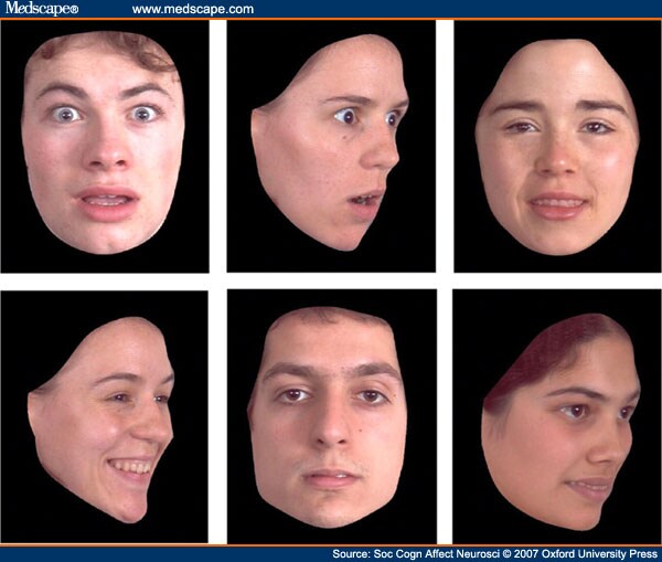


Figure 1. 

Examples of Face-stimuli; Fear Front 100%; Fear Side 100%; Happy Front 50%; Happy Side 100%; Neutral Front; Happy Side 50%.


There were five conditions: Happy-front, Happy-side, Fear-front, Fear-side and baseline. Stimuli were presented in the ratio of one-third 100% emotional expression, one-third 50% emotional expression and one-third neutral. Subjects were asked to decide if the emotional expression was neutral or emotional and then to press the appropriate button on their key-pad. In the baseline condition a cross was presented on the left or right of the screen and subjects were asked to press a button if the cross was on either the left or right of the screen.


Each condition consisted of 12 images in the proportions above, each presented for 2.5 s. Total block time was 32.5 s including 2.5 s for instruction. Each condition was repeated four times for a total session length of 10 min 50 s (5 conditions X 4 repeats X 32.5 sec). The session was repeated twice.


Scanning Method and Analysis


Functional imaging (FMRI) was performed using a 1.5 T scanner (NVi, General Electric Medical Systems, Milwaukee, WI). A quadrature head coil was used to obtain high-resolution gradient echo 3D volumetric images and four sets of functional images using blood oxygenation level dependent contrast. The high-resolution images were collected using a T1 weighted sequence with the following parameters: field of view, 24 cm; 20/6, (TR/TE); flip angle, 35°; slices, 124; slice thickness, 1.6 mm; and matrix, 256 X 256. FMR images were acquired in axial planes with a {T }2 *-weighted single shot, gradient-echo, echo-planar pulse sequence with the following parameters: field of view, 24 cm; 3000/33, (TR/TE); flip angle, 90°, slices, 24; slice thickness, 5 mm; and matrix, 128 X 128. The head was firmly stabilized between two foam pads.


Data analysis was performed in a two-stage mixed-effects analysis (equivalent to a random effects analysis) in which BOLD responses for each subject were first modeled using a standard hemodynamic function in the context of the fixed-effects general linear model. Subject-specific linear contrasts on the parameter estimates were then entered into a second-level analysis to perform within group analysis using a one-sample t-test, resulting in a t statistic for each voxel. For within group analyses, contrast weights were used to identify clusters of activation. The term 'activation' is defined as voxels showing significantly better fit of the hemodynamic model in one condition compared to another. For direct group comparison, two different weight combinations were used to pursue two kinds of contrast: T- group + 1/T+ group - 1 (to identify voxels showing significantly better fit of the hemodynamic model (i.e. 'greater activation') in the T- than in the T+ group); T+ group - 1/T- group + 1 (to identify voxels showing better fit for the T+ group).


Plasma Tryptophan Measures


The tryptophan assays were performed on the Biochrom 20 Plus amino acid analyser (from Biochrom Ltd, Cambridge, UK). This is a continuous flow liquid chromatography system utilizing cation exchange resin followed by treatment with ninhydrin solution. Plasma, treated with equal volumes of 0.1M HCl and 500 µmol/l norleucine in 10%(w/v) 5-sulphosalicylic acid, was mixed and centrifuged. The supernatant was stored frozen until a batch was collected. The thawed supernatants were treated with 0.50M LiOH and loaded onto the amino acid analyzer. Comparison to standard solutions allowed calculation of the tryptophan concentration.


Results


For each individual, sensitivity to ATD was calculated by measuring the plasma tryptophan level during the active session as a percentage of baseline level. Serum tryptophan was successfully lowered in all participants by up to 92% but the range was considerable (Supplementary Table 1 ). For two individuals, levels during the active session remained at 50-60% of baseline. Initial, group-wise, random effects analyses failed to reveal effects of tryptophan depletion on brain activity. After regression analysis (see subsequently) indicated a dose-dependent response to ATD, we deemed these two individuals to be non-responders and re-ran group-wise analyses of effects of ATD with them excluded. This meant that fMRI data was available from 10 participants (20 sessions) for fMRI analyses concerning the relationships between facial posture and emotional expression, and from eight participants for examining the interactions with tryptophan depletion. Reaction-time data was available for nine participants.


Main Effects


ATD. Our strategy was first to examine the main effects of ATD on whole brain analyses and then to explore interactions and regions of interest. We looked at overall effect of ATD on task by contrasting the sum of all emotional judgment conditions with baseline in the T+ and T- conditions separately, and then looking at where T+ activity was greater than T-. This revealed a focus of activity in left posterior superior temporal sulcus (STS) in addition to other activity. (Figure 2 and Supplementary Table 2 .)
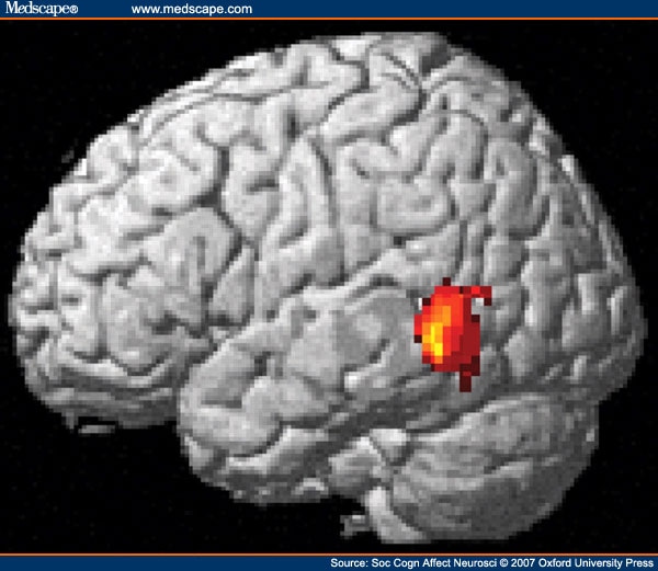


Figure 2. 

Main Effects of ATD on Task-related Activity in Left Posterior Superior Temporal Sulcus.


Face direction. Frontal viewing of faces was associated with higher activity compared to side-viewing in visual cortex, cingulate gyrus, premotor cortex and medial OFC (see Supplementary Material: Table 2 and Figure 1).


Emotion type. There were no main effects of emotion at the whole brain level.


Interactions


Emotion type and face-direction. An interaction between emotion and direction of expression was evident in a main cluster in the right parahippocampal gyrus and additional clusters in visual cortex (Supplementary Table 4).


ATD and face-direction. Interactions between ATD and the front-side contrast are shown in Figure 3 and Table 2 . There were five main areas: bilateral STS, the right TP, MPFC, left lateral OFC/dorsolateral prefrontal cortex and left amygdala. These clusters remained significant after correction for multiple comparisons.
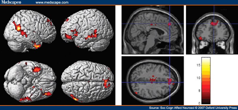


Figure 3. 

Interaction between the contrast of front vs side view of face and tryptophan non-depletion vs depletion. Top panel: rendering on cortical surface; Bottom panel: saggital section showing medial prefrontal cortex activity. Main foci are: bilateral along the length of the superior temporal sulcus, the right temporal pole, paracingulate region, left inferior frontal gyrus.


Local Analyses


To ascertain the changes that were driving these interactions, region of interest (ROI) analyses were conducted. ß-values were extracted for cubes of 27 voxels centered on peak voxels in clusters of interest. ATD had a marked impact on activity related to faces viewed from the front in MPFC, rTP, and OFC. Activity in these areas during side viewing of faces changed less with ATD than with front viewing of faces. For right STS and left Broca's area, effects of ATD were greater on side-viewing than front-viewing of faces. Activity that was greater for front compared to side before tryptophan depletion became greater for side compared to front after depletion. Interactions between ATD and emotion type were evident in the amygdala in whole brain analyses, but ROI analysis confined to the STS, MPFC and TPs failed to reveal an interaction between ATD and emotion in these areas. ROI analysis (Figure 4) in the amygdala shows that the interaction between ATD and emotion (x = -22, y = -1, z = -20: P = 0.036; x = 18, y = -7, z = -15: P = 0.040 following small volume correction) was driven through effects on happy as well as fear expressions. In the right amygdala, ATD led the amygdala to respond more strongly to side-happy expressions but had little effect on the response to fear. In the left amygdala, ATD affected the response to face-on-fear but not the response to happiness.
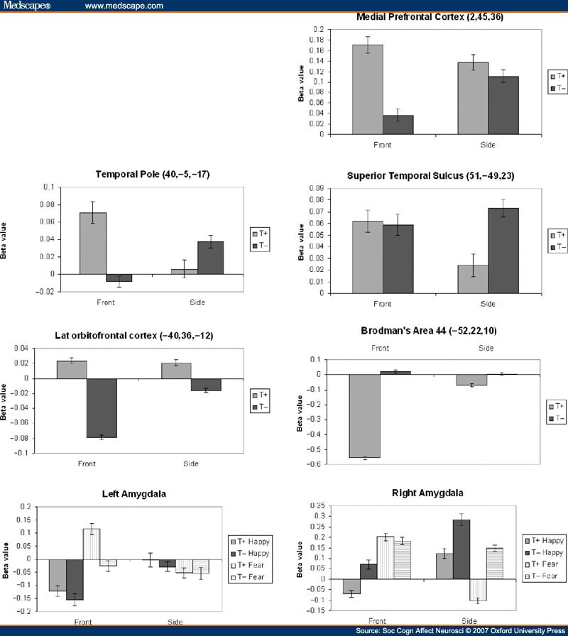


Figure 4. 

Graphs showing directions of change in activity following tryptophan depletion, driving interactions shown in Figure 1. Beta-values for 27 voxels surrounding peak value within each cluster, and reflect activity relative to average activity in that area over the duration of scans. Error bars constitute 95% confidence intervals.


Regression Analyses


We predicted individual variation in sensitivity to the ATD protocol, and hypothesized that serotonin function would be disrupted in a dose-dependent fashion in line with the drop in plasma levels of tryptophan. Therefore, in brain areas where serotonin production controlled activity levels, ATD sensitivity would correlate with the BOLD signal. We therefore performed a regression analysis to identify voxels where the mean differences for parameter estimates between active and baseline conditions correlated with plasma tryptophan reduction. We conducted analyses for all conditions together and then emotion types and face direction types separately. Results are shown in Figure 5, and Supplementary Tables 5-7. For all conditions, the largest clusters of correlated activity were located in hippocampus, and the intraparietal sulcus, extending from the angular gyrus along to the superior parietal lobule and precuneus. There were also significant clusters located in the left insula, caudal part of the anterior cingulate, left superior temporal gyrus (STG), and bilateral posterior temporo-occipital junctions. When analyses were separated according to emotion, the extent of depletion correlated more with activity for happy related expressions in anterior cingulate cortex. No correlations were found when analysis was carried out for separate facial directions.
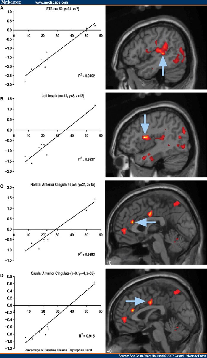


Figure 5. 

Areas of cortex where activity correlates strongly with sensitivity to tryptophan depletion. Graphs show serum tryptophan levels at scanning as percentage of baseline levels, plotted against activity averaged over whole cluster during viewing of faces, compared to baseline. Arrows point to peak of cluster. (A) All faces, peak z-score = 4.32, (B) All faces, peak z-score = 5.03 (C) Happy only, peak z-score = 5.15 (D) Happy only, peak z-score = 4.49. Full list of areas affected in supplementary tables 5–7.


Reaction Times


Overall, ATD was associated with a significantly faster response time (Response Time Mean ± 1SE: T- = 1.186 ± 0.076 T+ = 1.311 ± 0.068 s ANOVA main effect of ATD F1,8, = 8.6, P = 0.02). This is unlikely to be the result of a speed/accuracy trade-off because it was not associated with significantly greater numbers of errors (accuracy by mean errors ±1SE: T- = 20.2% ± 3.2% T+ = 14.1 ± 1.5%; ANOVA main effect of ATD non-significant F1,8, = 2.83, P = 0.13). However, ATD was associated with more errors when processing specifically emotions shown in profile, suggesting that ATD may have confounded the greater difficulty of judging emotions seen in profile (Accuracy by mean errors ± 1SE: T-front = 12.5% ± 3.8%, T+ front = 10.2 ± 1.3% T-side = 27.9% ± 3.3% T+ side = 18.0 ± 2.1%; ANOVA interaction between TDP and head view on accuracy judging fear and happiness: F1,8, = 7.89, P = 0.023).


Discussion


We predicted that associative learning and empathic functions serving emotional judgment would be more engaged when facial expression was directed to the observer, and so would be more susceptible to serotonin depletion. The interaction between ATD and facial direction provided support for this hypothesis. Clusters in lateral OFC and amygdala, areas associated with associative learning functions, showed that effects here were much more marked for front-viewed compared to side-viewed expressions. The same contrast also identified clusters in medial frontal cortex (MFC), temporal pole, insula and cingulate gyrus, as well as a dorsal prefrontal cluster that involved Broca's area. The cluster in the MFC maps closely onto an area associated with 'person-perception' (Iacoboni et al., 2004; Mason et al., 2004; Mitchell et al., 2005 Amodio and Frith, 2006) and falls at the boundary between anterior rostral MFC, thought to serve more emotional functions, and posterior rostral MFC thought to serve more cognitive functions (Steele and Lawrie, 2004). This might therefore justify the argument that the front-side X ATD interaction affected only emotional perception. However, areas associated with empathy were also identified in this interaction, including temporal pole (Carr et al., 2003; Frith and Frith, 2003; Kim et al., 2005; Amodio and Frith, 2006; Vollm et al., 2006), caudal cingulate (Lane et al., 1998; Singer et al., 2004), Broca's area and insula (Carr et al., 2003). Therefore, the interaction between ATD and direction of emotional expression is most consistent with an important role for serotonin in mediating the effects of an emotional expression on both associative learning and empathic function, which itself is dependent upon the face being viewed from the front. The regression analysis was also consistent with effects on empathic function, revealing effects of serotonin depletion on caudal anterior cingulate, though these were specific to processing happiness. Correlations may be greater for happiness than fear because of close connectivity with the amygdala, which could contribute more significantly to variance for fear, thus reducing the strength of the correlation. Reaction times were also consistent with effects of ATD on frontal lobe function. ATD resulted in faster response times in the absence of decreased accuracy for front-viewed faces, indicating that it favored more rapid decision making through diminished recruitment of higher cortical function to the task. The response time pattern became more complex when considering side vs front, presumably because of the greater difficulty of judging emotion at a lower impact for side-viewed faces.


Activity in STS was also affected bilaterally by ATD. On the left, this was independent of face view, whereas on the right, activity was increased for side-viewed expressions. Right STS may be more associated with attribution of intention than left STS (Saxe et al., 2004), suggesting that effects of ATD on right STS reflect decreased attention to emotional expression, but increased attention to the goal of gaze. In support of this view, we also found an interaction in BA 44, which is concerned with attributing the relationship between an action and its goal (Kakei et al., 2001; Ochiai et al., 2005). Effects in the amygdala were also lateralized and largely reflected changes in STS. On the right side, ATD increased activity for side-viewed faces, whilst on the left it was reduced or unchanged. There has been some debate with regards to the function of amygdala laterality. Here we suggest that the left is concerned with attention to gaze, whilst the right is more concerned with the goal of the gaze.


The adaptive function of these changes may be related to functions of serotonin in depression and anxiety. As mentioned earlier, it seems likely that ATD reduces serotonergic function, though there may be some question as to the exact mechanism by which this occurs. Reduced serotonin function is associated with greater anxiety and lowered mood, which may be associated with a more threatening and risky environment. In such environments it may be more adaptive to attend to external precipitants of emotional expressions, rather than the underlying mental states. In such cases, attention to the goal of gaze would be more adaptive than attention to the mental state underlying an emotional expression.


A limitation of this study was the low number of subjects in the final analysis of ATD effects. Nevertheless, our approach was robust, using a random effects analysis with a threshold set at P < 0.05 after adjustment for multiple comparisons. Our analyses are unlikely to have resulted in chance findings, though smaller effects may have gone undetected. Including our two non-responders in our regression analyses led to identification of close correlations between extent of tryptophan depletion and activity in anterior cingulate and paracingulate, as well as insula regions and left STS. Correlations are still evident in these regions with the non-responders removed but would not reach the level of significance required to emerge from a whole brain analysis.


To summarize our findings, we suggest that serotonin mediates the impact of a facial expression on emotion perception with marked consequences for empathic reactivity and emotional learning. Impaired serotonin function facilitates a shift of observer's attention to the goal of gaze rather than mental state associated with another's emotional expression. This may reflect adaptive changes to environmental modulators of mood, and may have implications for our understanding of disorders that involve dysfunctional serotonergic activity including depression (Caspi et al., 2003), autism (Devlin et al., 2005; McDougle et al., 2005) and psychopathy (Dolan and Anderson, 2003). Overall our work suggests a potentially powerful neurotransmitter based brain mechanism that could mediate between mental well-being and patterns of thinking that may heavily influence social behavior.


Table 1. Details of Stimuli Types


Front
Side
50% 100% 50% 100%
Neutral Neutral-front Neutral-side
Happy Happy-front (50%) Happy-front (100%) Happy-side (50%) Happy-side (100%)
Fear Fear-front (50%) Fear- front (100%) Fear-side (50%) Fear-side (100%)


Table 2. Detail of Clusters Shown in Figure 2. Paired T+ vs T-, Interaction With Front vs Side, Responders to ATD Only (n = 8). Loci Reported are Significant at 0.05 Level Corrected for Multiple Comparisons at Cluster Level (Location of Peak of Cluster in Bold, Other Loci are Sub-peaks Within Cluster, Reported to Indicate Direction of Clusters)


X Y Z Hemisphere Region BA Z Extent
40 -5 -17 Right Temporal Pole 20 5.08 717
36 -2 -35 Right Inferior Temporal Gyrus 20 3.84
40 1 -24 Right Middle Temporal Gyrus 21 3.78
-18 -6 -13 Left Amygdala - 4.45 781
-42 -10 -15 Left Sub-Gyral 20 4.34
-57 -22 -11 Left Middle Temporal Gyrus 21 4.13
51 -49 23 Right Supramarginal Gyrus 40 4.42 1004
40 -54 17 Right Superior Temporal Gyrus 22 4.07
48 -66 40 Right Inferior Parietal Lobule 39 4.00
2 45 36 Right Medial Prefrontal Cortex 6 4.12 488
18 49 42 Right Superior Frontal Gyrus 8 4.01
-8 34 28 Left Cingulate Gyrus 32 3.53
-28 7 -10 Left Subcallosal Gyrus 34 4.11 303
-50 22 10 Left Inferior Frontal Gyrus 45 3.72
-36 19 -6 Left Inferior Frontal Gyrus 47 3.05
-40 36 -12 Left Middle Frontal Gyrus 11 3.67 273
-38 48 -16 Left Middle Frontal Gyrus 11 3.11
-44 52 -6 Left Middle Frontal Gyrus 10 3.09
-12 -6 44 Left Cingulate Gyrus 24 3.65 283
-16 -12 36 Left Cingulate Gyrus 24 2.94
-4 -10 39 Left Cingulate Gyrus 24 2.89
-10 -39 2 Left Parahippocampal Gyrus 30 3.61 272
-8 -39 -5 Left Parahippocampal Gyrus 30 3.11
42 -7 11 Right Insula 13 3.56 299
40 0 6 Right Insula 13 3.34
28 -1 13 Right Lentiform Nucleus - 3.19


References


  • Adams R.B. Jr, Gordon HL, Baird AA, Ambady N, Kleck RE. Effects of gaze on amygdala sensitivity to anger and fear faces. Science (2003) 300:1536.

  • Adams R.B. Jr, Kleck RE. Perceived gaze direction and the processing of facial displays of emotion. Psychological Science (2003) 14:644-7.

  • Adolphs R, Gosselin F, Buchanan TW, Tranel D, Schyns P, Damasio AR. A mechanism for impaired fear recognition after amygdala damage. Nature (2005) 433:68-72.

  • Amodio DM, Frith CD. Meeting of minds: the medial frontal cortex and social cognition. Nature Reviews Neuroscience (2006) 7:268-77.

  • Attenburrow MJ, Williams C, Odontiadis J, Reed A, Powell J, Cowen PJ, Harmer CJ. Acute administration of nutritionally sourced tryptophan increases fear recognition. Journal of Psychopharmacology (2003) 169:104-07.

  • Baron-Cohen S, Ring HA, Wheelwright S, et al. Social intelligence in the normal and autistic brain: an fMRI study. European Journal of Neuroscience (1999) 11:1891-8.

  • Bediou B, Franck N, Saoud M, et al. Effects of emotion and identity on facial affect processing in schizophrenia. Psychiatry Research (2005) 133:149-57.

  • Blair RJ, Morris JS, Frith CD, Perrett DI, Dolan RJ. Dissociable neural responses to facial expressions of sadness and anger. Brain (1999) 122:883-93.

  • Blair RJR, Mitchell DGV, Peschardt KS, et al. Reduced sensitivity to others' fearful expressions in psychopathic individuals. Personality and Individual Differences (2004) 37:1111-22.

  • Calder AJ, Young AW, Rowland D, Perrett DI, Hodges JR, Etcoff NL. Facial emotion recognition after bilateral amygdala damage: Differentially severe impairment of fear. Cognitive Neuropsychology (1996) 13:699-745.

  • Calder AJ, Young AW, Rowland D, Perrett DI. Computer-enhanced emotion in facial expressions. (1997) Proceedings of the Royal Society of London, 264. 919-25.

  • Canli T, Omura K, Haas BW, Fallgatter A, Constable RT, Lesch KP. Beyond affect: a role for genetic variation of the serotonin transporter in neural activation during a cognitive attention task. (2005) Proceedings of the National Academy of Sciences, USA, 102. 12224-9.

  • Carr L, Iacoboni M, Dubeau M-C, Mazziota JC, Lenzi GL. Neural mechanisms of empathy in humans: a relay from neural systems for imitation to limbic areas. (2003) Proceedings of the National Academy of Sciences, USA, 100. 5497-502.

  • Caspi A, Sugden K, Moffitt TE, et al. Influence of life stress on depression: moderation by a polymorphism in the 5-HTT gene. Science (2003) 301:386-9.

  • Celada P, Puig MV, Casanovas JM, Guillazo G, Artigas F. Control of dorsal raphe serotonergic neurons by the medial prefrontal cortex: involvement of serotonin-1A, GABA(A), and glutamate receptors. Journal of Neuroscience (2001) 21:9917-29.

  • Cools R, Calder AJ, Lawrence AD, Clark L, Bullmore E, Robbins TW. Individual differences in threat sensitivity predict serotonergic modulation of amygdala response to fearful faces. Psychopharmacology (2005) 180:670-9.

  • Dalton KM, Nacewicz BM, Johnstone T, et al. Gaze fixation and the neural circuitry of face processing in autism. Nature Neuroscience (2005) 8:519-26.

  • Devlin B, Cook EH Jr, Coon H, et al. Autism and the serotonin transporter: the long and short of it. Molecular Psychiatry (2005) 10:1110-16.

  • Dolan MC, Anderson IM. The relationship between serotonergic function and the Psychopathy Checklist: Screening Version. Journal of Psychopharmacology (2003) 17:216-22.

  • Evers EAT, van der Veen FM, van Deursen JA, Schmitt JAJ, Deutz NEP, Jolles J. The effect of acute tryptophan depletion on the BOLD response during performance monitoring and response inhibition in healthy male volunteers. Psychopharmacology (2006) 187:200-8.

  • Frigerio E, Burt DM, Montagne B, Murray LK, Perrett DI. Facial affect perception in alcoholics. Psychiatry Research (2002) 113:161-71.

  • Frith U, Frith CD. Development and neurophysiology of mentalizing. Philosophical Transactions of the Royal Society of London Series B-Biological Sciences (2003) 358:459-473.

  • Gallese V, Keysers C, Rizzolatti G. A unifying view of the basis of social cognition. Trends in Cognitive Sciences (2004) 8:396-403.

  • Hariri AR, Mattay VS, Tessitore A, et al. Serotonin transporter genetic variation and the response of the human amygdala. Science (2002) 297:400-3.

  • Harmer CJ, Mackay CE, Reid CB, Cowen PJ, Goodwin GM. Antidepressant drug treatment modifies the neural processing of nonconscious threat cues. Biological Psychiatry (2006) 59:816-20.

  • Harmer CJ, Rogers RD, Tunbridge E, Cowen PJ, Goodwin GM. Tryptophan depletion decreases the recognition of fear in female volunteers. Psychopharmacology (2003) 167:411-7.

  • Hietanen JK, Surakka V, Linnankoski I. Facial electromyographic responses to vocal affect expressions. Psychophysiology (1998) 35:530-6.

  • Iacoboni M, Lieberman MD, Knowlton BJ, et al. Watching social interactions produces dorsomedial prefrontal and medial parietal BOLD fMRI signal increases compared to a resting baseline. NeuroImage (2004) 21:1167-73.

  • Kakei S, Hoffman DS, Strick PL. Direction of action is represented in the ventral premotor cortex. Nature Neuroscience (2001) 4:1020-5.

  • Kennedy SH, Evans KR, Kruger S, et al. Changes in regional brain glucose metabolism measured with positron emission tomography after paroxetine treatment of major depression. American Journal of Psychiatry (2001) 158:899-905.

  • Kim JW, Kim JJ, Jeong BS, et al. Neural mechanism for judging the appropriateness of facial affect. Brain Research and Cognitive Brain Research (2005) 25:659-67.

  • Kringelbach ML, Rolls ET. The functional neuroanatomy of the human orbitofrontal cortex: evidence from neuroimaging and neuropsychology. Progress in Neurobiology (2004) 72:341-372.

  • Lane RD, Reiman EM, Axelrod B, Yun LS, Holmes A, Schwartz GE. Neural correlates of levels of emotional awareness: evidence of an interaction between emotion and attention in the anterior cingulate cortex. Journal of Cognitive Neuroscience (1998) 10:525-35.

  • Lawrence K, Campbell R, Swettenham J, et al. Interpreting gaze in Turner syndrome: impaired sensitivity to intention and emotion, but preservation of social cueing. Neuropsychologia (2003) 41:894-905.

  • Lembke A, Ketter TA. Impaired recognition of facial emotion in mania. American Journal of Psychiatry (2002) 159:302-4.

  • Lord C, Risi S, Lambrecht L, et al. The autism diagnostic observation schedule-generic: a standard measure of social and communication deficits associated with the spectrum of autism. Journal of Autism and Developmental Disorders (2000) 30:205-223.

  • Mason MF, Banfield JF, Macrae CN. Thinking about actions: the neural substrates of person knowledge. Cerebral Cortex (2004) 14:209-14.

  • McDougle CJ, Erickson CA, Stigler KA, Posey DJ. Neurochemistry in the pathophysiology of autism. Journal of Clinical Psychiatry (2005) 66:9-18.

  • Milders M, Crawford JR, Lamb A, Simpson SA. Differential deficits in expression recognition in gene-carriers and patients with Huntington's disease. Neuropsychologia (2003) 41:1484-92.

  • Mitchell JP, Macrae CN, Banaji MR. Forming impressions of people versus inanimate objects: Social-cognitive processing in the medial prefrontal cortex. NeuroImage (2005) 26:251-7.

  • Morris JS, Frith CD, Perrett DI, et al. A differential neural response in the human amygdala to fearful and happy facial expressions. Nature (1996) 383:812-5.

  • Ochiai T, Mushiake H, Tanji J. Involvement of the ventral premotor cortex in controlling image motion of the hand during performance of a target-capturing task. Cerebral Cortex (2005) 15:929-37.

  • Phelps EA. Emotion and cognition: insights from studies of the human amygdala. Annual Review of Psychology (2006) 57:27-53.

  • Phillips ML, Young AW, Senior C, et al. A specific neural substrate for perceiving facial expressions of disgust. Nature (1997) 389:495-8.

  • Robbins TW. Chemistry of the mind: Neurochemical modulation of prefrontal cortical function. Journal of Comparative Neurology (2005) 493:140-6.

  • Rubinow DR, Post RM. Impaired recognition of affect in facial expression in depressed patients. Biological Psychiatry (1992) 31:947-53.

  • Sato W, Yoshikawa S, Kochiyama T, Matsumura M. The amygdala processes the emotional significance of facial expressions: an fMRI investigation using the interaction between expression and face direction. NeuroImage (2004) 22:1006-13.

  • Saxe R, Xiao DK, Kovacs G, Perrett DI, Kanwisher N. A region of right posterior superior temporal sulcus responds to observed intentional actions. Neuropsychologia (2004) 42:1435-46.

  • Singer T, Seymour B, O'Doherty J, Kaube H, Dolan RJ, Frith CD. Empathy for pain involves the affective but not sensory components of pain. Science (2004) 303:1157-62.

  • Steele JD, Lawrie SM. Segregation of cognitive and emotional function in the prefrontal cortex: a stereotactic meta-analysis. NeuroImage (2004) 21:868-75.

  • Surakka V, Hietanen JK. Facial and emotional reactions to Duchenne and non-Duchenne smiles. International Journal of Psychophysiology (1998) 29:23-33.

  • Vollm BA, Taylor AN, Richardson P, et al. Neuronal correlates of theory of mind and empathy: a functional magnetic resonance imaging study in a nonverbal task. NeuroImage (2006) 29:90-8.

  • Wicker B, Perrett DI, Baron-Cohen S, Decety J. Being the target of another's emotion: a PET study. Neuropsychologia (2003) 41:139-46.

  • Williams JHG. Directedness, egocentrism, & autism. In: An Integrated View of Autism: Perspectives from Neurocognitive, Clinical and Intervention Research.—McGregor E, Nunez M, Williams K, Gomez JC, eds. (2007) Oxford, UK: Blackwells. (In press).

  • Yatham LN, Liddle PF, Shiah IS, et al. Effects of rapid tryptophan depletion on brain 5-HT(2) receptors: a PET study. British Journal of Psychiatry (2001) 178:448-53.

  • Young AW, Rowland D, Calder AJ, Etcoff NL, Seth A, Perrett DI. Facial expression megamix: tests of dimensional and category accounts of emotional recognition. Cognition (1997) 63:271-313.

  • Young SN, Smith SE, Pihl RO, Ervin FR. Tryptophan depletion causes a rapid lowering of mood in normal males. Psychopharmacology (1985) 87:173-77.


Acknowledgements

We are grateful to our volunteers, Sophia Durrani for stimulus preparation, and Bill Mutch in the Biochemistry Department of Aberdeen Royal Infirmary for tryptophan measures.


Funding Information

The project was funded by the National Alliance for Autism Research.


Reprint Address

Correspondence to: Dr Justin H. G. Williams, Department of Child Health, University of Aberdeen Medical School, Royal Aberdeen Children's Hospital, Aberdeen. AB25 2ZG. E-mail: justin.williams@abdn.ac.uk


Justin H. G. Williams,* David I. Perrett,1 Gordon D. Waiter,2 Stephen Pechey3

*Department of Child Health, University of Aberdeen Medical School, Aberdeen, Scotland, UK,
1School of Psychology, University of St Andrews, St Andrews, Scotland, UK,
2Department of Radiology, University of Aberdeen, Aberdeen, Scotland, UK, and
3School of Psychology, University of Central Lancashire, Preston, England, UK


Disclosure: None declared.

Tags: ,
Powered by Qumana


Neurobiology of Depression: An Integrated View Of Key Findings

V. Maletic; M. Robinson; T. Oakes; S. Iyengar; S. G. Ball; J. RussellInt J Clin Pract.  2007;61(12):2030-2040.  ©2007 Blackwell Publishing

Posted 12/18/2007


Summary


Aims: The objectives of the present review were to summarise the key findings from the clinical literature regarding the neurobiology of major depressive disorder (MDD) and their implications for maximising treatment outcomes. Several neuroanatomical structures in the prefrontal and limbic areas of the brain are involved in affective regulation. In patients with MDD, alterations in the dynamic patterns of activity among these structures have profound implications for the pathogenesis of this illness.
Discussion: The present work reviews the evidence for the progressive nature of MDD along with associated changes in neuroanatomical structure and function, especially for the hippocampus. The role of glucocorticoids, inflammatory cytokines and brain-derived growth factors are discussed as mediators of these pathological alterations. From this integrated model, the role of antidepressant therapy in restoring normative processes is examined along with additional treatment guidelines.
Conclusion: Major depressive disorder is an illness with significant neurobiological consequences involving structural, functional and molecular alterations in several areas of the brain. Antidepressant pharmacotherapy is associated with restoration of the underlying physiology. Clinicians are advised to intervene with MDD using an early, comprehensive treatment approach that has remission as the goal.


Review Criteria


The search strategies used for this review involved literature searches of the MEDLINE and Psychinfo electronic databases. The main heading terms included major depressive disorder, neurobiology, antidepressant, hippocampus, brain-derived neurotrophic growth factor, glucocorticoids and monoamines. As part of the research strategy, each article's bibliography was reviewed for additional potential research findings relevant to these terms.


Message for the Clinic


Major depressive disorder (MDD) is an illness with significant neurobiological consequences involving structural, functional and molecular alterations in several areas of the brain. Antidepressant pharmacotherapy is associated with restoration of the underlying physiology. Clinicians are advised to intervene with MDD using an early, comprehensive treatment approach that has remission as the goal.


Introduction


Major depressive disorder (MDD) remains one of the most frequently seen psychiatric illnesses in primary care settings.[1] Although family and primary care physicians have greatly increased their recognition and treatment of this illness, MDD remains an unresolved treatment challenge for many physicians and patients.[2] Increasing evidence has accrued in recent years regarding the impact of MDD on the structural and functional processes occurring in the brain. From the initial views that depression was caused by 'chemical imbalance' in the brain, this body of research has developed into a complex theory involving neuronal networks and plasticity.[3] The network model has also led to a greater understanding of the mechanisms of effective treatment interventions and their role in mitigating the deleterious effects of MDD.[4]


The objectives of the present review were to summarise the key findings from the clinical literature regarding the neurobiology of MDD and their implications for maximising treatment outcomes. First, the evidence that MDD is not only a chronic and recurrent illness, but also a progressive illness will be presented. Second, the impact of MDD on the primary neuroanatomical sites associated with mood regulation will be described at the structural and functional level. Third, the molecular processes that have been implicated for mediating these structural and functional changes will be explored. Fourth, the role of multiple neurotransmitter systems will be reviewed for their involvement in restoration and recovery from MDD. The last section will discuss the treatment guidelines for obtaining remission in the context of this neurobiological model.


Major Depressive Disorder as a Progressive Illness


Epidemiological studies have consistently shown that MDD is one of the most prevalent lifetime psychiatric disorders. In the National Comorbidity Replication Survey, based on DSM-IV criteria for MDD, the lifetime prevalence rate was 16.2%, with a 12-month estimate of 6.6%.[5] The presentation of MDD is heterogeneous with respect to both core and associated symptoms.[6] In the Diagnostic and Statistical Manual of Mental Disorders Fourth Edition, Text Revision,[7] the diagnosis of MDD requires the experience of major depressive episodes that are defined by at least five of the following symptoms for at least 2 weeks duration: loss of interest, depressed mood, appetite/weight disturbance, sleep disturbance, psychomotor change, loss of energy, worthlessness/guilt, concentration difficulties/indecisiveness and thoughts of death/suicide. Depressed mood or loss of interest must be one of the symptoms, but with the inclusion of compound criteria (e.g. worthlessness or guilt), a diagnosis of MDD can be met by various permutations, and episodes may then be further qualified by other associated features (e.g. postpartum, seasonal pattern, with melancholy or psychotic symptoms).


Even though MDD is characterised as an episodic illness, prospective studies have found that recurrence is the norm rather than the exception. For example, in a naturalistic, 15-year follow-up of a sample of 380 patients experiencing an index MDD episode, 73% experienced a recurrent episode,[8] with each subsequent episode increasing the probability of further episodes.[9] Similarly, in the STAR*D Project (Sequenced Treatment Alternatives to Relieve Depression) that includes 1500 patients with MDD, 74% of patients had experienced more than one episode.[10] Recurrence of MDD appears to be driven in part by neurobiological vulnerabilities. In the STAR*D Project, patients who experienced multiple episodes were more likely to have a positive family history of depressive illness and an earlier age of onset of their index depressive episode compared with patients who were in their first episode.[10]


Consistent evidence has also supported a 'kindling hypothesis' in which depressive episodes become more easily triggered over time.[11] As the number of depressive episodes increase, future episodes are predicted more by the number of prior episodes rather than by life stress.[12] Figure 1 Kindling can be described as a process which occurs by a lowering of the threshold for the impact of stressful life events (i.e. sensitisation to minor events) or by an increase in spontaneous dysregulation, both of which could indicate progressive effects of MDD.[13] An analysis of the risk of recurrence in a large study of twins also suggests genetic contributions as patients with a high genetic risk were 'prekindled'; that is, they had a lower association between stressful life events and the onset of depressive episodes compared with patients having a low genetic risk.[14]


Figure 1. 

Major depression as a progressive illness. As the number of major depressive episodes increase, the risk for subsequent episodes is predicted more from the number of prior episodes and less from the occurrence of a recent life stress. Figure adapted from ref. no.[14]


Early adverse experiences may also contribute to long-term neurobiological alterations associated with depression. In preclinical studies, maternal deprivation of rat pups during critical development periods resulted in subsequent hyper-reactivity to stress and marked behavioural changes in adult rats.[15] In children who had a history of early maltreatment, the risk for depressive symptoms was associated with an interaction between genotypes [e.g. serotonin (5-HT) transporter] and history of maltreatment.[16] Considering these findings, some researchers have suggested that greater neurobiological changes occur in patients with depression who have early adverse experiences compared with patients who are depressed but do not have such a history, indicating that these patients may represent an especially vulnerable subtype of depressive illness.[17]


Chronicity also suggests long-term neurobiological consequences associated with the MDD illness. In the STAR*D Project, 25% of the patients (with single or recurrent MDD) were identified as having a chronic episode of more than 2 years duration.[10] In another large multicentre treatment study (n = 681), patients' depression was classified using DSM-IV modifiers into four categories: chronic MDD (episodes > 2 years), MDD with incomplete recovery (partial response), MDD superimposed on dysthymia (double depression) and chronic MDD superimposed on dysthymia (depressive symptoms > 4 years). Despite multiple comparisons across a broad range of clinical and psychological variables, few differences were found among the four groups, resulting in the conclusion that various manifestations of chronic depression represent the same illness.[18]


As the duration of depressive episodes increases, the probability of recovery substantially decreases over time. In a 5-year prospective study of outpatients with depression, approximately half recovered within the first 6 months, but afterwards the rate of recovery diminished substantially. For example, patients who had experienced depressive episodes of 1-year duration had a recovery rate of 16% compared with a 1% recovery rate for patients whose episodes persisted > 5 years.[19] Similarly, in a prospective study of new onset depressive episodes, a longer duration (> 12 weeks) of previous episodes reduced the likelihood of recovery from the new onset episode by 37%.[20]


Even if patients no longer meet full criteria for an MDD episode, studies have found that a substantial subset of patients continue to experience residual symptoms and diminished functioning. In a 3-year longitudinal epidemiological study, 165 patients were assessed before and after an MDD episode. Although mean values on functional measures returned to premorbid levels, 15-40% of patients experienced a worsening in psychosocial functioning that persisted after the episode, and the overall functioning of the entire sample continued to be lower than that of a healthy cohort.[21] In a 10-year, naturalistic longitudinal study, patients who experienced subthreshold depressive symptoms following an MDD episode were at significantly greater risk for a recurrence, and they also had a much faster onset of their next episode compared with patients whose episode had fully remitted, suggesting that residual symptoms represent vulnerability because of an active disease state.[22]


The recurrence and chronicity of MDD along with possible kindling effects have shifted the perspective of the appropriate treatment goal. The gold standard for treatment outcome has been raised from response (reduction in symptoms) to remission (absence of symptoms) or recovery (extended period of remission).[23] However, obtaining recovery implies not only the remission of symptoms but also a restoration of the underlying physiology associated with the illness. Therefore, further understanding of the neurobiological changes associated with MDD is necessary for identifying true recovery processes.


Functional and Structural Changes in MDD


Although much information still needs to be attained, imaging and other methods have begun to elucidate the neurobiological abnormalities associated with MDD. In particular, several prefrontal and limbic structures and their interconnected circuits have been implicated in affective regulation. Figure 2 These neuroanatomical areas include the ventromedial prefrontal cortex (VMPFC), lateral orbital prefrontal cortex (LOPFC), dorsolateral prefrontal cortex (DLPFC), anterior cingulated cortex (ACC), ventral striatum (including nucleus accumbens), amygdala and the hippocampus. Abnormalities in these areas have been shown in patients with MDD compared with healthy controls and thus suggest a foundation for the symptomatic expression of MDD.[24,25] However, these deviations may be obscured or not present at the individual patient level and thus, these findings cannot necessarily be considered pathognomic.


Figure 2. 

Major depressive disorder affects the dynamic connectivity among neuroanatomical structures involved in regulation of mood and stress response. Limbic structures (amygdala, hippocampus and nucleus accumbens) have reciprocal connections with 'para-limbic´ cortical areas, subgenual anterior cingluate and ventromedial prefrontal cortex (VMPFC). Hypothetically, disrupted 'connectivity´ between limbic/para-limbic areas and rostral integrative prefrontal formations, results in compromised feedback regulation of limbic activity. Consequently, dorsal cognitive/executive network is hypoactive while overly active limbic areas continue to stimulate the hypothalamus leading to neuroendocrine dysregulation and sympathetic hyperactivity


As an integrated circuit, the prefrontal cortex, cingulate, amygdala, and hippocampus serves not only mood regulation, but also learning and contextual memory processes. Within the prefrontal cortex, the VMPFC mediates pain, aggression, sexual functioning and eating behaviours whereas the LOPFC assesses risk and modulates maladaptive and perseverative affective states (behaviours). These two areas have a reciprocal pattern of activity with the DLFPC, which maintains executive function, effortful sustained attention, and working memory processes.[26] Subdivisions within the ACC assume diverse roles, with the dorsal ACC being part of the cognitive/executive functioning network and the ventral ACC being involved in assessing emotional and motivational information. The ACC also monitors outcomes of behaviour and cognition and makes adjustments based on changing contingencies.[27,28]


In patients with MDD, regional blood flow studies suggest hyperactivity in the VMPFC and LOPFC and hypoactivity in the DLFPC compared with controls.[24] Given the functions of these regions, as previously described, this abnormal activity pattern may be responsible for the manifestations of symptoms associated with MDD. Hyperactivity of the VMPFC is associated with enhanced sensitivity to pain, anxiety, depressive ruminations and tension whereas hypoactivity of the DLFPC may produce psychomotor retardation, apathy, and deficits in attention and working memory. Using fMRI paradigms, connectivity studies have also suggested a decrement in the 'communication' between amygdala and ACC regions.[29] A consequence of this loss of connectivity could be a failure of the ACC to serve its inhibitory role in emotional regulation,[30] resulting in further motivational and affective disruption.[31]


At the intersection of limbic, cognitive/executive and neuroendocrine regulatory circuits, including the hypothalamic-pituitary-adrenal axis (HPA), the hippocampus may be particularly vulnerable in depression. Imaging studies of hippocampal volume have been of particular interest. In a meta-analysis of 12 studies, hippocampal volume was found to be consistently and significantly reduced in patients with MDD compared with controls, and these reductions occurred bilaterally with a slightly greater decrement in right hippocampal volume.[32] Other studies have shown that the degree of hippocampal reduction is directly proportional to the number and the duration of untreated depressive episodes.[33] Among depressed inpatients, while controlling for the effect of age, hippocampal volume was significantly correlated with duration of illness prior to hospitalisation.[34] Even after remission of an episode,patients with recurrent MDD have continued to show significantly smaller hippocampal volume compared with healthy controls.[35]


Differences in hippocampal volume between patients with depression and healthy controls may not be fully attributable to the disease state. Heritability studies of hippocampal volume suggest both environmental and genetic contributions with heritability estimates of 54% in nonhuman primates and 40% in adult male twins.[36,37] Several genomic imaging studies, comparing patients with MDD and healthy controls, have shown associations between hippocampal volume and specific genes that are implicated in mood disorders.[38,39] In a 1-year prospective study of 30 patients with MDD, hippocampal volume did not significantly change during the study period, but patients whose depression failed to remit had a significantly smaller hippocampus at baseline and at 1 year than did patients who did remit.[40] Combining the evidence from these genetic, cross-sectional, and clinical treatment studies suggests that morphological differences in the hippocampus may be a predisposing factor in MDD, but changes can also accumulate in the course of the disease and thereby create an obstacle to full recovery.


Molecular Processes Mediating Neurobiological Changes


The alteration in the hippocampus signifies a potential outcome of injurious feedback that occurs via neuroendocrine dysregulation. A consistent finding in patients with MDD is a high level of the stress hormone cortisol, which may cause impairment in neuroplasticity and cellular resistance.[41] An imbalance between glucocorticoid and mineral corticoid receptors in MDD along with high-density glucocorticoid receptors (GRs) may also contribute to the hippocampus' susceptibility to neuronal damage.[42] Subsequent hippocampal atrophy could result in further neuroendocrine dysfunction and hence a potential 'run-away' system.[43] Postmortem comparisons of brain tissue in patients with MDD and age-matched healthy controls have shown hippocampal shrinkage in depressed subjects that was caused by increased density of neuronal cells and a significant reduction in neuropil (i.e. decreased dendridic branching and spine complexities).[44]


A corollary of elevated glucocorticoids and compromised hippocampal functioning may also be the down-regulation of the GR sensitivity. Under conditions of chronic stress, decrease in GR sensitivity can have negative consequences as GR signalling becomes insufficient to 'turn off' the initial responses to stress as part of a negative feedback process.[45,46] Figure 3 Subsequently, HPA hypothalamic overactivity, in conjunction with amygdala activation, leads to increased sympathetic tone, which promotes the release of cytokines from macrophages. Increase in pro-inflammatory cytokines has been associated with loss of insulin and GR sensitivity, which further perpetuates metabolic and neuroendocrine disruption.[47] Symptomatically, disruptions as a result of proinflammatory cytokines may be experienced as fatigue, loss of appetite and libido as well as hypersensitivity to pain.[48]


Figure 3. 

Molecular processes are impacted by stress and depression. Stress results in release of glucocorticoids and corticotrophin releasing hormones (CRH) and pro-inflammatory cytokines (TNF, IL-1, IL-6). In depression, disruption of serotonin (5-HT), norepinephrine (NE) and dopamine (DA) transmission impair the regulatory feedback loops that 'turn off´ the stress response. Sympathetic overactivity contributes to immune activation and release of inflammatory cytokines. Inflammatory cytokines further interfere with monoaminergic and neurotrophic signalling. They may also diminish central corticosteroid receptor sensitivity, leading to disruption of feedback control. Figure adapted from ref. no.[46]


Proinflammatory cytokines may also diminish neurotrophic support and monoamine neurotransmission that can lead to neuronal apoptosis and glial damage. Alterations in glia-neuron relationships have been recently emphasised in the aetiology of neuropathic pain and MDD.[47,49] Glia cells are involved in an intricate interaction with neurons in which astroglia and microglia maintain homeostasis of the neuronal environment by modulating electrolytes, neurotransmitters, cytokines and neurotrophic factors.[50] Neurons reciprocate support of glial function via neurotrophin signalling. Stress, depression and ensuing peripheral immune dysregulation lead to activation of microglia that then contribute to the existing immune disruption by additional release of inflammatory cytokines.[51]


An integral part of maintaining the health of these glial-neuron interactions may be mediated by brain-derived neurotrophic factor (BDNF).[52] Involved in neurogenesis, BDNF is the primary neurotrophin of the hippocampus. As a dimeric protein involved in cell maintenance, plasticity, growth and death (apoptosis), BDNF is structurally related to nerve growth factor and is distributed widely throughout the brain.[53] When BDNF interacts with tyrosine receptor kinase receptors (TRkB), it promotes cellular resilience and long-term potentiation. However, the precursor form of BDNF (pro-BDNF) can also precipitate reduction in dendritic spines and cell death when it binds with the p75 receptor. Thus, depending upon its expression, BDNF can prune neural networks in an activity dependent manner that is regulated by various neurotransmitters [glutamate, GABA, 5-HT, norepinephrine (NE), acetylcholine, dopamine and hormones].[54]


Preclinical and clinical studies have suggested dysregulation in BDNF occurs under conditions of chronic stress and depression. In animal models, acute and chronic immobilisation stress resulted in decreased BDNF expression using mRNA assays. Similar results were also observed following administration of acute and chronic pain stimuli.[55] Within humans, levels of serum BDNF has been found to be significantly lower in untreated patients with MDD compared with treated patients or healthy controls.[56] Similarly, postmortem analyses of brains of persons who committed suicide showed that BDNF and another neurotrophin (NT-3) were significantly reduced compared with non-suicide controls.[57]


From the above observations, the neurotrophic hypothesis has emerged as a major theory for the pathogenesis of major depression. In this model, stress and genetic vulnerability elevate glucocorticoid steroids and alter cellular plasticity via downregulation of growth factors and receptor sensitivity.[4] The reduction in growth factors, such as BDNF, impacts negatively on the structural and functional processes within the limbic system, especially for the hippocampus. Chronic and recurrent MDD may result in subsequent atrophy and further disruptions in neurocircuitry. From this hypothesis, recovery and remission of MDD would be dependent upon a reversal of these processes, such as an increase in BDNF levels.


Complementing the neurotrophic hypothesis of MDD is the monoamine theory, which postulates that depression is associated with low levels of monoamines, particularly, 5-HT and NE. A recent imaging study of patients with untreated depression found a high global receptor density for the monoamine oxidase A (MAO-A), which nonspecifically metabolises these neurotransmitters. In this updated theory, long-term monoamine loss because of this global MAO-A activity interacts with regional specific transporter densities (i.e. 5-HT, NE), resulting in the expression of the depressive illness.[58] Both 5-HT and NE ascending fibres originate from brainstem nuclei and innervate the limbic system, prefrontal cortex and associated structures involved in the regulation of mood. Descending pathways project through the dorsolateral spinal column and are instrumental in the regulation of pain.[59,60] Therefore, depending upon the specific transporter densities within these regions, various symptoms of depression (mood, cognition and pain) will be manifested within the context of the overall global reduction in monoamine levels.[58]


Role of Neurotransmitters in Recovery From MDD


Therapeutically, selective serotonergic reuptake inhibitors (SSRIs) and NE reuptake inhibitors (NRIs) are known to increase their respective monoamine levels in the brain. Chronic treatment with monoamine reuptake inhibitors increases activation of cyclic adenosine 3-5 monophosphatase (cAMP), which in turn stimulates protein kinase A. Activation of this protein enzyme regulates target genes leading to an increase in BDNF synthesis.[52] The antidepressant-induced cAMP activity can also enhance GR sensitivity and inhibit cytokine signalling, further assisting in the restoration of the neurocircuitry feedback loops.[61]


The effect of increasing monoamine levels (dopamine, 5-HT and NE) on BDNF and growth factors may be one mechanism that produces the antidepressant response. Preclinical study of rat brain cells has demonstrated that monoamenergic activity (NE, 5-HT) upregulates BDNF synthesis in astrocytes.[62] Clinically, successful treatment with antidepressants results in normalisation of serum BDNF level, which is considered an indirect measure of cortical BDNF activity. Support for the relationship between serum and cortical BDNF levels has been derived from correlations in animal studies as well as findings that serum BDNF passes the blood-brain barrier and reflects stored and circulating BDNF in humans.[63,64] In a study of 10 patients who were treated for 12 weeks with a dual reuptake inhibitor, improvement in depressive symptoms was correlated with increases in BDNF levels, and the BDNF levels of remitted patients had normalised to the same level observed in healthy controls.[65] Response to various SSRI and 5-HT noradrenalin reuptake inhibitors (SNRI) treatments has been similarly associated with restoration of normative BDNF values.[66] Figure 4 Postmortem analysis of brain tissue has shown that subjects who had been treated with an antidepressant at time of death had greater hippocampal BDNF expression as measured by immunoreactivity than did untreated subjects with mood disorders.[67]


Figure 4. 

Antidepressant therapy is associated with restoring normative processes. Treatment with various selective serotonin antidepressant treatments and serotonergic noradrenergic reuptake inhibitors resulted in increases in serum brain-derived neurotrophic factor (BDNF) for patients with MDD to levels comparable that were observed with healthy controls. Reprinted with copyright permission from ref. no.[66]


Antidepressant therapeutic response is also associated with re-establishment of normative cortical activity. A study of 17 inpatients with MDD examined regional activity changes following 1 week and 6 week fluoxetine treatment. At 1 week, all patients showed increases in hippocampal activity and decreases in posterior cingulate and prefrontal cortex activity. After 6 weeks of treatment, patients who had responded to treatment showed a reversal of this pattern with decreased limbic activity and increased prefrontal cortical activity whereas non-responders continued to show the 1-week pattern.[68] Normalisation in the amygdala and ACC has also been associated with positive response to treatment. Using a masking paradigm for subconscious activation, patients with MDD showed a baseline hyper-reactivity of the left amygdala that attenuated following 8-week treatment with sertraline.[69]


Other lines of evidence also support the restorative nature of antidepressant therapy. Structural and functional MRI assessments of patients with MDD who were treated with fluoxetine indicated the importance of ACC grey matter volume for treatment response as there was a positive association among grey matter volume, normalisation of ACC activity, and response to treatment.[70] Conversely, in patients with MDD who failed to respond to antidepressant treatment, plasma levels of proinflammatory cytokines were elevated compared with healthy controls or euthymic patients with MDD.[71]


Symptomatically, improvements in specific MDD symptoms have been associated with regional improvements in brain metabolic activity. In 39 outpatients with MDD, improvement in cognitive symptoms was correlated with increases in DLPFC and improvements in fatigue/psychomotor retardation was associated with decreases in VMPFC activity. Interestingly, these changes were seen in responders regardless of whether treatment was pharmacological or psychological.[72] Restoration of the neurobiological regulation in MDD via neurotrophic factors and neurogenesis appears to be a common factor across various effective treatments for MDD, including pharmacological, psychological and somatic treatments, such as diet and exercise.[73]


Treatment Implications of the Neurobiological Model


The neurobiological sequelae and repercussions of chronic or recurrent MDD indicate that interventions for MDD should be focused on achieving optimal treatment early. Longitudinal studies have shown that one of the best predictors of remission status at 2 years was response to acute treatment, i.e. initial 6 weeks.[74] In addition, the adequacy of treatment may also have prognostic implications. For patients with late-life depression, exposure to previous inadequate trials of antidepressants resulted in a reduced response rate to pharmacological intervention augmented by psychotherapy compared with treatment of naive patients, even after controlling for baseline severity.[75] Similarly, in a large observational study of 996 patients with MDD, non-response or incomplete response to initial antidepressant treatment was a significant predictor of eventual treatment resistance.[76] On the positive side, an early response to antidepressants has been shown to predict greater treatment adherence.[77]


One way of maximising early response is to apply a comprehensive treatment that increases activity of multiple monoaminergic systems. In a double-blind, randomised treatment study, 39 inpatients with MDD received either fluoxetine (a serotonergic intervention), desipramine (a noradrenergic intervention) or their combination. After 6 weeks of treatment, patients who had been given the combination treatment were more likely to achieve remission (53.8%) than either intervention alone (0 % and 7.1%).[78] Similarly, a recent large meta-analysis encompassing 93 trials and 17,036 patients compared efficacy outcomes of SSRI with SNRI treatments for MDD that showed a modest but significant advantage in efficacy with SNRI treatments.[79] An earlier meta-analysis did not find a difference in efficacy between SSRIs and dual acting agents (mostly tricyclic antidepressants), with the exception of the inpatient populations, where dual acting tricyclic antidepressants had an advantage.[80] Thus, although current treatment algorithms for MDD usually are initiated with SSRIs, the role of combination treatment or dual reuptake inhibitors are increasingly being considered as a preferred option.[81]


Another advantage of targeting both of 5-HT and NE systems is improvement not only in the core features of MDD, but also in associated physical symptoms. Painful physical symptoms are prevalent in patients with MDD, and these symptoms increase the illness burden and impair the ability to attain remission.[82,83] In a study of primary care patients with MDD who were treated with SSRIs for 9 months, mood symptoms continued to improve over time while painful physical symptoms persisted.[84] The occurrence of painful physical symptoms and MDD reflects the shared underlying pathophysiology between mood and pain regulation. Importantly, there may be also a synergistic interaction between the 5-HT and NE systems to obtain analgesia. In an animal model of pain, treatment with dual reuptake inhibitors or combination treatment (5-HT/NE) appeared to enhance the effectiveness of pain alleviation.[85] Clinically, patients with MDD who experienced a 50% or greater reduction in pain were more likely to achieve remission than patients whose pain reduction was < 50%.[86]


With remission and recovery as the goal, the treatment guidelines derived from the neurobiological model emphasise the need for not only early and comprehensive intervention, but also vigorous attention to residual symptoms. In a 2-year study of outpatients with MDD, patients who obtained only a partial remission of symptoms were more likely to relapse (67.5%) than patients who had attained full remission (15.2%).[87] Specific recommendations for the treatment of residual symptoms have not been determined empirically, but likely require additional augmentation with other pharmacological and psychological treatments; in addition to reducing the risk of relapse, the treatment of residual symptoms may enhance compliance and long-term outcomes.[88]


Conclusions


As the underlying neurobiological model of depression is increasingly understood, treatment providers are directed to recognise that the factors that may initiate a MDD episode and those that maintain the illness are likely to be very different. Genetic and stress vulnerabilities interplay to initiate a cascade of neurobiological alterations that disrupt a dynamic system. Progressive effects of recurrent and chronic MDD may then be potentiated by further structural and functional abnormalities.


Given these long-term consequences, an essential objective of treatment must be to restore normative functioning and prevent further neurobiological structural alterations. Increasing 5-HT and NE neurotransmission is likely to initiate true recovery with the restoration of neurotrophic support, glucocorticoid signalling and neuroendocrine regulation. The use of dual reuptake inhibitors enhances the probability of remission as it addresses the complex interplay of the emotional and physical symptoms of MDD. Painful physical symptoms are increasingly recognised as having a significant impact on functioning and recovery; thus, affirming the need for antidepressant treatments that can effectively reduce these symptoms as well.


From the neurobiological model, the treatment guidelines of early, comprehensive and progressive treatment require a change in perspective for both patients and providers. A residual symptom may be interpreted as a proxy of an active disease state, with ensuing structural alterations and systemic consequences. With remission and recovery as the goal, patients will need to be educated about the benefits of long-term treatment rather than episodic or incomplete intervention. A biopsychosocial treatment model that incorporates cognitive-behavioural or interpersonal therapy along with pharmacological interventions serves to address both the initiation and maintenance factors and can reduce the risk of relapse.[89] Once remission is attained, maintenance of effect may become the more appropriate term, rather than relapse prevention, to emphasise the necessity for an ongoing collaboration between patient and physician in order to maintain neurobiological homeostasis.


CLICK HERE for subscription information about this journal.


References


  1. Remick RA. Diagnosis and management of depression in primary care: a clinical update and review. Can Med Assoc J 2002; 167: 1253–60.

  2. Nemeroff CB. The burden of severe depression: a review of diagnostic challenges and treatment alternatives. J Psychiatr Res 2007; 41: 189–206.

  3. Castren E. Is mood chemistry? Nat Rev Neurosci 2005; 6: 241–6.

  4. Duman RS, Monteggia LM. A neurotrophic model for stress-related mood disorders. Biol Psychiatry 2006; 59: 1116–27.

  5. Kessler RC, Berglund P, Demler O et al. The epidemiology of major depressive disorder: results from the National Comorbidity Survey Replication (NCS-R). JAMA 2003; 289: 3095–105.

  6. McGlinchey JB, Zimmerman M, Young D, Chelminski I. Diagnosing major depressive disorder VII: are some symptoms better than others? J Nerv Ment Dis 2006; 194: 785–90.

  7. American Psychiatric Association. Diagnostic and Statistical Manual for Mental Disorders-Fourth Edition, Text-Revision. Washington, DC: American Psychiatric Press, 2000.

  8. Mueller TI, Leon AC, Keller MB et al. Recurrence after recovery from major depressive disorder during 15 years of observational follow-up. Am J Psychiatry 1999; 156: 1000–6.

  9. Solomon DA, Keller MB, Leon AC et al. Multiple recurrences of major depressive disorder. Am J Psychiatry 2000; 157: 229–33.

  10. Hollon SD, Shelton RC, Wisniewski S et al. Presenting characteristics of depressed outpatients as a function of recurrence: preliminary findings from the STAR*D clinical trial. J Psychiatr Res 2006; 40: 59–69.

  11. Post RM, Rubinow DR, Ballenger JC. Conditioning and sensitisation in the longitudinal course of affective illness. Br J Psychiatry 1986; 149: 191–201.

  12. Kendler KS, Thornton LM, Gardner CO. Stressful life events and previous episodes in the etiology of major depression in women: an evaluation of the "kindling" hypothesis. Am J Psychiatry 2000; 157: 1243–51.

  13. Monroe SM, Harkness KL. Life stress, the ''kindling'' hypothesis, and the recurrence of depression: considerations from a life stress perspective. Psychol Rev 2005; 112: 417–45.

  14. Kendler KS, Thornton LM, Gardner CO. Genetic risk, number of previous depressive episodes, and stressful life events in predicting the onset of major depression. Am J Psychiatry 2001; 158: 582–6.

  15. Ladd CO, Huot RL, Thrivkraman KV, Nemeroff CB, Meany MJ, Plotsky PM. Long-term behavioral and neuroendocrine adaptations to adverse early experience. Prog Brain Res 2000; 122: 81–103.

  16. Kaufman J, Yang BZ, Douglas-Palumberi H et al. Brain-derived neurotrophic factor-5-HTTLPR gene interactions and environmental modifiers of depression in children. Biol Psychiatry 2006; 59: 673–80.

  17. Heim C, Plotsky PM, Nemeroff CB. Importance of studying the contributions of early adverse experience to neurobiological findings in depression. Neuropsychopharmacology 2004; 29: 641–8.

  18. McCullough JP Jr, Klein DN, Borian FE et al. Group comparisons of DSM-IV subtypes of chronic depression: validity of the distinctions, part 2. J Abnorm Psychol 2003; 112: 614–22.

  19. Keller MB, Lavori PW, Mueller TI et al. Time to recovery, chronicity, and levels of psychopathology in major depression. A 5-year prospective follow-up of 431 subjects. Arch Gen Psychiatry 1992; 49: 809–16.

  20. Spijker J, de Graaf R, Bijl RV et al. Determinants of persistence of major depressive episodes in the general population. Results from the Netherlands Mental Health Survey and Incidence Study (NEMESIS). J Affect Disord 2004; 81: 231–40.

  21. Buist-Bouwman MA, Ormel J, de Graaf R, Vollebergh WAM. Functioning after a major depressive episode: complete or incomplete recovery? J Affect Disord 2004; 82: 363–71.

  22. Judd LL, Akiskal HS, Maser JD et al. Major depressive disorder: a prospective study of residual subthreshold depressive symptoms as predictor of rapid relapse. J Affect Disord 1998; 50: 97–108.

  23. Keller MB. Past, present, and future directions for defining optimal treatment outcome in depression: remission and beyond. JAMA 2003; 289: 3152–60.

  24. Drevets WC. Functional neuroimaging studies of depression: the anatomy of melancholia. Annu Rev Med 1998; 49: 341–61.

  25. Davidson RJ. Affective neuroscience and psychophysiology: toward a synthesis. Psychophysiology 2003; 40: 655–65.

  26. Swanson LW. The hypothalamus. In: Björklund A, Hökfelt T, Swanson LW, eds. Handbook of Chemical Neuroanatomy, Vol. 5, Integrated Systems of the CNS, Part I, Hypothalamus, Hippocampus, Amygdala, Retina. New York, NY: Elsevier, 1987: 1–124.

  27. Bush G, Luu P, Posner MI. Cognitive and emotional influences in anterior cingulate cortex. Trends Cogn Sci 2000; 4: 215–22.

  28. McCormick LM, Ziebell S, Nopoulos P et al. Anterior cingulated cortex: an MRI-based parcellation method. Neuroimage 2006; 32: 1167–75.

  29. Anand A, Li Y, Wang Y et al. Activity and connectivity of brain mood regulating circuit in depression: a functional magnetic resonance study. Biol Psychiatry 2005; 57: 1079–88.

  30. Whittle S, Allen NB, Lubman DI, Yücel M. The neurobiological basis of temperament: towards a better understanding of psychopathology. Neurosci Biobehav Rev 2005; 30: 511–25.

  31. MacDonald AW III, Cohen JD, Stenger VA, Carter CS. Dissociating the role of the dorsolateral prefrontal and anterior cingulate cortex in cognitive control. Science 2000; 288: 1835–8.

  32. Videbech P, Ravnkilde B. Hippocampal volume and depression: a meta-analysis of MRI studies. Am J Psychiatry 2004; 161: 1957–66.

  33. Sheline YI, Gado MH, Kraemer HC. Untreated depression and hippocampal volume loss. Am J Psychiatry 2003; 160: 1516–8.

  34. Colla M, Kronenberg G, Deuschle M et al. Hippocampal volume reduction and HPA-system activity in major depression. J Psychiatr Res 2007; 41: 553–60.

  35. Neumeister A, Wood S, Bonne O et al. Reduced hippocampal volume in unmedicated, remitted patients with major depression versus control subjects. Biol Psychiatry 2005; 57: 935–7.

  36. Lyons DM, Yang C, Sawyer-Glover AM, Moseley ME, Schatzberg AF. Early life stress and inherited variation in monkey hippocampal volumes. Arch Gen Psychiatry 2001; 58: 1145–51.

  37. Sullilvan EV, Pfefferbaum A, Swan GE, Carmelli D. Heritability of hippocampal size in elderly twin men: equivalent influence from genes and environment. Hippocampus 2001; 11: 754–62.

  38. Frodl T, Meisenzahl EM, Zill P et al. Reduced hippocampal volumes associated with the long variant of the serotonin transporter polymorphism in major depression. Arch Gen Psychiatry 2004; 61: 177–83.

  39. Frodl T, Schüle C, Schmitt G et al. Association of the brain-derived neurotrophic factor Val66Met polymorphism with reduced hippocampal volumes in major depression. Arch Gen Psychiatry 2007; 64: 410–6.

  40. Frodl T, Meisenzahl EM, Zetzsche T et al. Hippocampal and amygdala changes in patients with major depressive disorder and healthy controls during a 1-year follow-up. J Clin Psychiatry 2004; 65: 492–9.

  41. Manji HK, Quiroz JA, Sporn J et al. Enhancing neuronal plasticity and cellular resilience to develop novel, improved therapeutics for difficult-to-treat depression. Biol Psychiatry 2003; 53: 707–42.

  42. de Kloet ER, Derijk RH, Meijer OC. Therapy insight: is there an imbalanced response of mineralcorticoid and glucocorticoid receptors in depression? Nat Clin Pract Endocrinol Metab 2007; 3: 168–79.

  43. Nestler EJ, Barrot M, DiLeone RJ et al. Neurobiology of depression. Neuron 2002; 34: 13–25.

  44. Stockmeier CA, Mahajan GJ, Konick LC et al. Cellular changes in the postmortem hippocampus in major depression. Biol Psychiatry 2004; 56: 640–50.

  45. Raison CL, Miller AH. When not enough is too much: the role of insufficient glucocorticoid signaling in the pathophysiology of stress-related disorders. Am J Psychiatry 2003; 160: 1554–65.

  46. Raison CL, Capuron L, Miller AH. Cytokines sing the blues: inflammation and the pathogenesis of depression. Trends Immunol 2006; 27: 24–31.

  47. Weisler-Frank J, Maier SF, Watkins LR. Immune-to-brain communication dynamically modulates pain: physiological and pathological consequences. Brain Behav Immun 2005; 19: 104–11.

  48. Tsigos C, Chrousos GP. Hypothalamic-pituitary-adrenal axis, neuroendocrine factors, and stress. J Psychosom Res 2002; 53: 865–71.

  49. Rajkowska G, Miguel-Hidalgo JJ. Gliogenesis and glial pathology in depression. CNS Neurol Disord Drug Targets 2007; 6: 219–33.

  50. Bessis A, Bechade C, Bernard D, Roumier A. Microglial control of neuronal death and synaptic properties. Glia 2007; 55: 233–8.

  51. Frank MG, Baratta MV, Sprunger DB, Watkins LR, Maier SF. Microglia serve as a neuroimmune substrate for stress-induced potentiation of CNS pro-inflammatory cytokine responses. Brain Behav Immun 2007; 21: 47–59.

  52. Duman RS, Heninger GR, Nestler EJ. A molecular and cellular theory of depression. Arch Gen Psychiatry 1997; 54: 597–606.

  53. Tapia-Arancibia L, Rage F, Givalois L, Arancibia S. Physiology of BDNF: focus on hypothalamic function. Front Neuroendocrinol 2004; 25: 77–107.

  54. Lu B, Pang PT, Woo NH. The yin and yang of neurotrophin action. Nat Rev Neurosci 2005; 6: 603–14.

  55. Duric V, McCarson KE. Hippocampal neurokinin-1 receptor and brain-derived neurotrophic factor gene expression is decreased in rat models of pain and stress. Neuroscience 2005; 133: 999–1006.

  56. Shimizu E, Hashimoto K, Okamura N et al. Alterations of serum levels of brain-derived neurotrophic factor (BDNF) in depressed patients with or without antidepressants. Biol Psychiatry 2003; 54: 70–5.

  57. Karege F, Vaudan G, Schwald M et al. Neurotrophin levels in postmortem brains of suicide victims and the effects of antemortem diagnosis and psychotropic drugs. Brain Res Mol Brain Res 2005; 136: 29–37.

  58. Meyer JH, Ginovart N, Boovariwala A et al. Elevated monoamine oxidase A levels in the brain: an explanation for the monoamine imbalance of major depression. Arch Gen Psychiatry 2006; 63: 1209–16.

  59. Mega MS, Cummings JL, Salloway S, Malloy P. The limbic system: an anatomic, phylogenetic, and clinical perspective. J Neuropsychiatry Clin Neurosci 1997; 9: 315–30.

  60. Fields HL, Heinricher MM, Mason P. Neurotransmitters in nociceptive modulatory circuits. Annu Rev Neurosci 1991; 14: 219–45.

  61. Pace TWW, Hu F, Miller AH. Cytokine-effects on glucocorticoid receptor function: relevance to glucocorticoid resistance and the pathophysiology and treatment of major depression. Brain Behav Immun 2007; 21: 9–19.

  62. Juric DM, Miklic S, Carman-Krzan M. Monominergic neuronal activity up-regulates BDNF synthesis in cultured neonatal ratastrocytes. Brain Res 2006; 1108: 54–62.

  63. Karege F, Schwald M, Cisse M. Postnatal development profile of brain-derived neurotrophic factor in rat brain and platelets. Neurosci Lett 2002; 328: 261–4.

  64. Lommatzsch M, Zingler D, Schuhbaeck K et al. The impact of age, weight, and gender on BDNF levels in human platelets and plasma. Neurobiol Aging 2005; 26: 115–23.

  65. Aydemir O, Deveci A, Taneli F. The effect of chronic antidepressant treatment on serum brain-derived neurotrophic factor levels in depressed patients: a preliminary study. Prog Neuropsychopharmacol Biol Psychiatry 2005; 29: 261–5.

  66. Gonul AS, Akdeniz F, Taneli F et al. Effect of treatment on serum brain-derived neurotrophic factor levels in depressed patients. Eur Arch Psychiatry Clin Neurosci 2005; 255: 381–6.

  67. Chen B, Dowlatshahi D, MacQueen GM et al. Increased hippocampal BDNF immunoreactivity in subjects treated with antidepressant medication. Biol Psychiatry 2001; 50: 260–5.

  68. Mayberg HS, Brannan SK, Tekell JL et al. Regional metabolic effects of fluoxetine in major depression: serial changes and relationship to clinical response. Biol Psychiatry 2000; 48: 830–43.

  69. Sheline YI, Barch DM, Donnelly JM et al. Increased amygdala response to masked emotional faces in depressed subjects resolves with antidepressant treatment: an fMRI study. Biol Psychiatry 2001; 50: 651–8.

  70. Chen CH, Ridler K, Suckling J et al. Brain imaging correlates of depressive symptom severity and predictors of symptom improvement after antidepressant treatment. Biol Psychiatry 2007; 62: 407–14.

  71. O'Brien SM, Scully P, Fitzgerald P et al. Plasma cytokine profiles in depressed patients who fail to respond to selective serotonin reuptake inhibitor therapy. J Psychiatr Res 2007; 41: 326–31.

  72. Brody AL, Saxena S, Mandelken MA et al. Brain metabolic changes associated with symptom factor improvement in major depressive disorder. Biol Psychiatry 2001; 50: 171–8.

  73. Duman RS. Neurotrophic factors and regulation of mood: role of exercise, diet, and metabolism. Neurobiol Aging 2005; 26 (Suppl. 1): S88–93.

  74. Szádóczky E, Rozsa S, Zambori J, Furedi J. Predictors for 2 year outcome of major depressive episode. J Affect Disord 2004; 83: 49–57.

  75. Tew JD, Mulsant BH, Houck PR et al. Impact of prior treatment exposure on response to antidepressant treatment in late life. Am J Geriatr Psychiatry 2006; 14: 957–65.

  76. Oswald P, Souery D, Kasper S et al. Predictive factors of resistance to antidepressant treatment: results from a European multicentre study. Eur Neuropsychopharmacol 2005; 15 (Suppl. 3): S326–7.

  77. Brook OH, van Hout HPJ, Stalman WAB, de Haan M. Nontricylic antidepressants: predictors of nonadherence. J Clin Psychopharmacol 2006; 26: 643–7.

  78. Nelson JC, Mazure CM, Jatlow PI et al. Combining norepinephrine and serotonin reuptake inhibition mechanism for treatment of depression: a double-blind, randomized study. Biol Psychiatry 2004; 55: 296–300.

  79. Papakostas GI, Thase ME, Fava M, Nelson JC, Shelton RC. Are antidepressant drugs that combine serotonergic and noradrenergic mechanisms of action more effective than the selective serotonin reuptake inhibitors in treating major depressive disorder? A meta-analysis of studies of newer agents. Biol Psychiatry 2007; doi: 10.1016/j.biopsych2007.03.027.

  80. Anderson I. Selective serotonin reuptake inhibitors versus tricyclic antidepressants: a meta-analysis of efficacy and tolerability. J Affect Dis 2000; 58: 19–36.

  81. Trivedi MH, Fava M, Marangell LB et al. Use of treatment algorithms for depression. Prim Care Companion J Clin Psychiatry 2006; 8: 291–7.

  82. Bair MJ, Robinson RL, Katon W, Kroenke K. Depression and pain comorbidity: a literature review. Arch Intern Med 2003; 163: 2433–45.

  83. Karp JF, Scott J, Houck P et al. Pain predicts longer time to remission during treatment of recurrent depression. J Clin Psychiatry 2005; 66: 591–7.

  84. Greco T, Eckert G, Kroenke K. The outcomes of physical symptoms with treatment of depression. J Gen Intern Med 2004; 19: 813–8.

  85. Jones CK, Eastwood BJ, Need AB, Shannon HE. Analgesic effects of serotonergic, noradrenergic or dual reuptake inhibitors in the carrageenan test in rats: evidence for synergism between serotonergic and noradrenergic reuptake inhibitors. Neuropharmacology 2006; 51: 1172–80.

  86. Fava M, Mallinckrodt CH, Detke MJ et al. The effect of duloxetine on painful physical symptoms in depressed patients: do improvements in these symptoms result in higher remission rates? J Clin Psychiatry 2004; 65: 521–30.

  87. Pintor L, Gastó C, Navarro V et al. Relapse of major depression after complete and partial remission during a 2-year follow-up. J Affect Disord 2003; 73: 237–44.

  88. Menza M, Marin H, Opper RS. Residual symptoms in depression: can treatment be symptom-specific? J Clin Psychiatry 2003; 64: 516–23.

  89. Paykel ES, Scott J, Cornwall PL et al. Duration of relapse prevention after cognitive therapy in residual depression: follow-up of controlled trial. Psychol Med 2005; 35: 59–68.


Disclaimer

Re-use of this article is permitted in accordance with the Creative Commons Deed, Attribution 2.5, which does not permit commercial exploitation.


Reprint Address

Vladimir Maletic, School of Medicine, University of South Carolina, 38 Parkway Commons Way, Greer, SC 29650, USA Tel.: + 864 848 4448 Fax: + 864 848 4428 Email: vmaletic@bellsouth.net


V. Maletic,1 M. Robinson,2 T. Oakes,2 S. Iyengar,2 S. G. Ball,2,3 J. Russell2

1School of Medicine, University of South Carolina, Greer, SC, USA,
2Eli Lilly and Company, Indianapolis, IN, USA,
3Indiana University School of Medicine, Indianapolis, IN, USA


Disclosure: Vladimir Maletic has served on the Speaker's Bureau or has been a consultant for Eli Lilly and Company and Cephalon. He did not receive any financial compensation for his work on this manuscript. His co-authors are each employees and/or shareholders of Eli Lilly and Company.


Tags: ,
Powered by Qumana


Medscape Psychiatry

Scientific American - Mind & Brain

Nature Reviews Neuroscience - Issue - nature.com science feeds

Nature Reviews Neuroscience - AOP - nature.com science feeds

Nature Neuroscience - Issue - nature.com science feeds

Nature Neuroscience - AOP - nature.com science feeds

Translational Psychiatry

Neuropsychopharmacology - AOP - nature.com science feeds

Neuropsychopharmacology - Issue - nature.com science feeds